One of the quiet scientific revolutions of the last 30 years has been in our ability to efficiently create materials with defined properties on demand and to exacting specifications. When you add non-carbon elements to artificial diamond, they form various “defect systems”. Our research uses one such defect, the nitrogen-vacancy (NV) centre. This defect emits light and can also be used as a sensor of electric and magnetic fields. We aim to combine “super-resolution” microscopes with the imaging of NV centres to measure biological fields with exquisite sensitivity and accuracy. The dream is to view the firing of neural cells in real time and with a resolution and field of view that could encompass networks of tens or hundreds of neurons. This will allow the detailed mapping of biological structures and, since we can use adaptive optics techniques to correct for any optical aberrations, these structures can be located in living tissue.
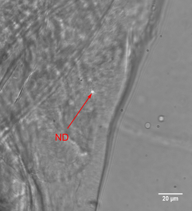
We are aiming to use small particles of diamond, nanodiamond, to get the defect centres as close to the source of the biological fields as possible. Apart from their potential for field sensing, the ability to have super-resolution compatible labels that do not photobleach and are biologically compatible and inert offers some interesting avenues for future research.